
457 Lake Cook Road (Deerfield Park Plaza)
Deerfield, IL 60015
Email Us:
Email us directly [+]
Fax: (847) 291-9362
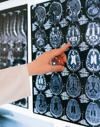
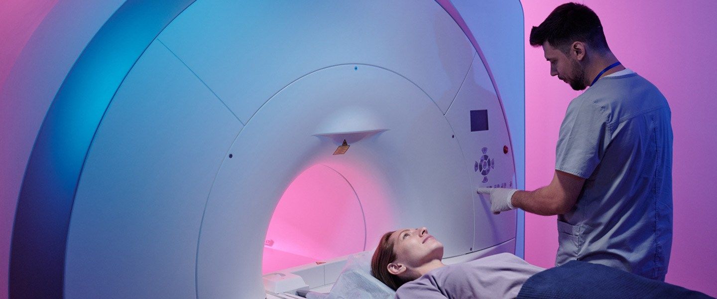
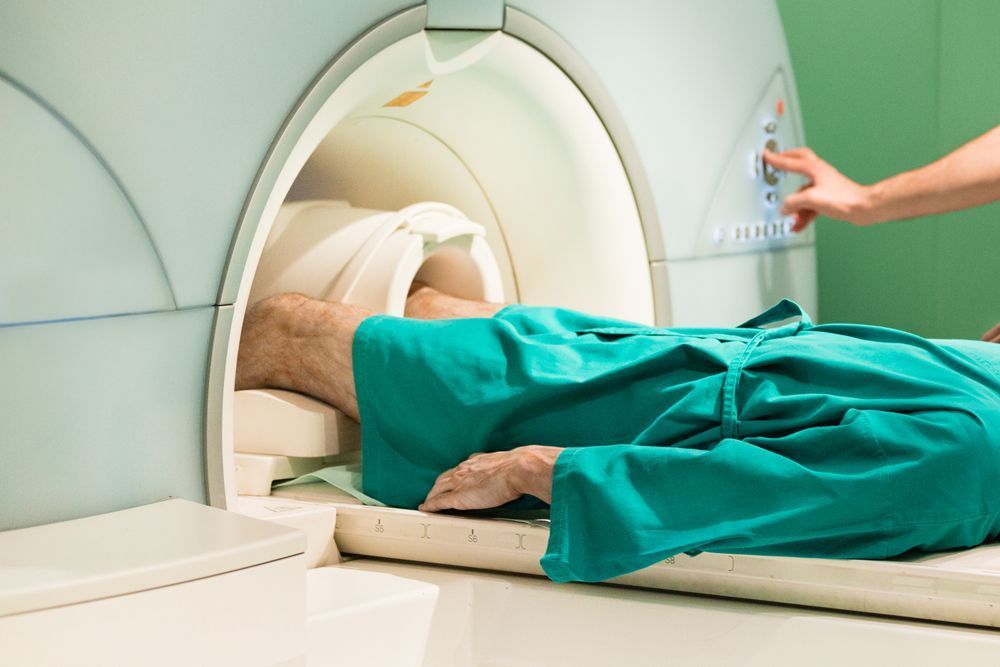
9 Conditions that the Upright MRI Can Detect in The Body and Brain
If you experience strange headaches, have a broken bone, or need to gain clarity about anything happening inside your body, your doctor may have you get a magnetic resonance imaging (MRI) scan to diagnose a disease, fracture or condition.
MRIs can detect a wide arrange of issues and conditions from head to toe. Here are some of the most common:
1. Tumors
You might have heard this word being thrown around before regarding cancer, but might not know what it actually is. Tumors are a big cluster of abnormal cells that eventually cause a swelling and may look like a lump from the outside perspective. Tumors can be benign (non-cancerous) or cancerous, so it’s imperative to get them scanned and diagnosed if you think you may have a tumor.
The Open Upright MRI can diagnose tumors in the brain or any other part of the body by offering patients a multi-position body scan.

2. Multiple Sclerosis
Multiple sclerosis (MS) is a disease that attacks the nervous system (brain and spinal cord) and disrupts the flow of information from the brain to the rest of the body. This results in symptoms like difficulty walking, talking, muscle spasms, and fatigue.
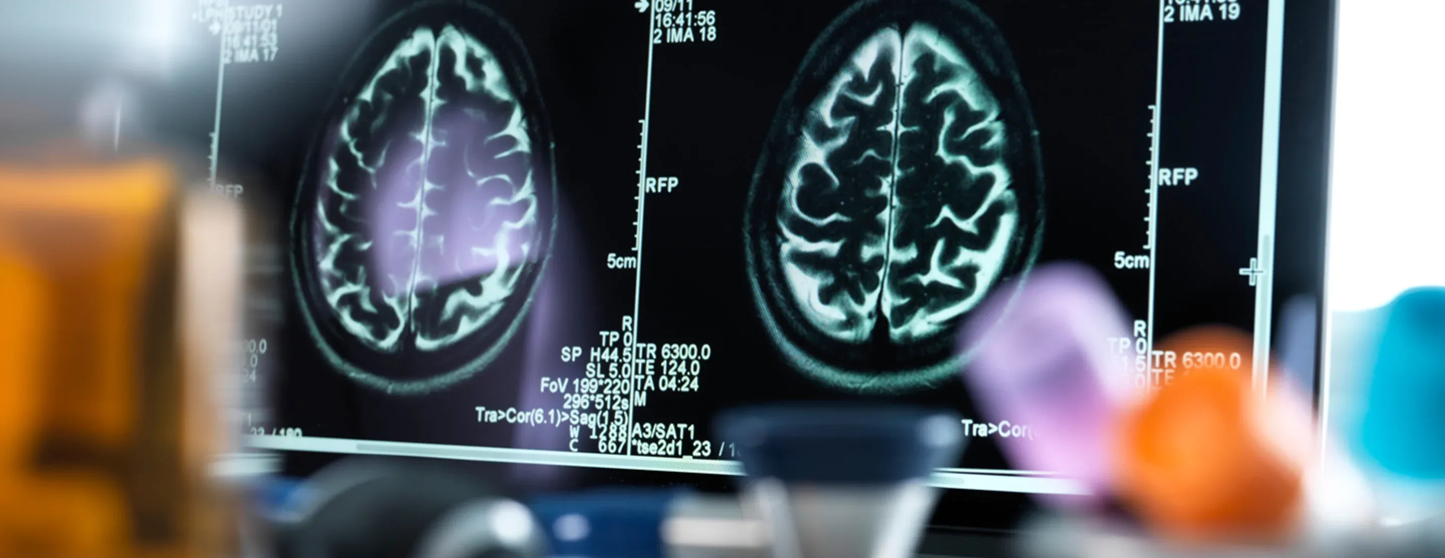
The recent technological advances from the FONAR Open Upright MRI have dramatically improved our understanding and diagnostic abilities of MS. Patients who may have MS receive a brain MRI, where physicians can identify lesions in the brain that show up in the MRI images as white or dark spots.
The lesions may or may not be MS, and having MS doesn’t necessarily mean you have lesions. In fact, according to National MS Society, MRI shows there are no lesions in 5 percent of patients with “clinically definite MS” at the time of diagnosis.
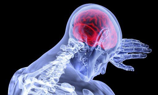
3. Stroke
Strokes occur when the blood supply in arteries that connect to the brain is blocked or reduced, which results in essentially an attack on the brain. When someone has a stroke, they may have a sudden weakness, trouble walking, talking, and even seeing. It happens extremely fast and can be deadly, so it is critical to get medical attentions as soon as possible.
MRIs have the ability to show areas of the brain in great detail and are used for patients who have suffered a stroke to analyze the damage and treatment options. The image that MRIs produce can show the region of the brain that was damaged, which helps physicians determine next steps in treatment.
4. Dementia
Dementia is a very common group of conditions associated with aging that cause memory loss, anxiety and paranoia. The most common cause of dementia is Alzheimer’s, a disease where brain cell connections and cells themselves die off and destroy memory. There is no cure for Alzheimer’s yet, but medications and treatment can help manage symptoms.
While no diagnostic test alone can diagnose dementia, MRI scans are used for Alzheimer’s or other dementias to track the change of the disease over time. Because of its ability to track changes, MRI has been pivotal in dementia research.
5. Traumatic Brain Injury
A traumatic brain injury, or TBI, is usually caused by a violent blow or penetration to the head and causes brain disarray. According to Medline Plus, some common causes for these severe head injuries include falls, sports injuries, child abuse, being struck by an object, or being hit or shot with a weapon.
A newer technique using contrast agents has helped physicians visualize the damage of a TBI or concussion. Essentially, a contrast agent is a dye that can be injected or consumed before the MRI scan that makes the images from the MRI clearer.
At Upright MRI of Deerfield, a contrast agent may be necessary in order to make a proper diagnosis. In our experience, 5 to 10 percent of patients require a contrast agent.
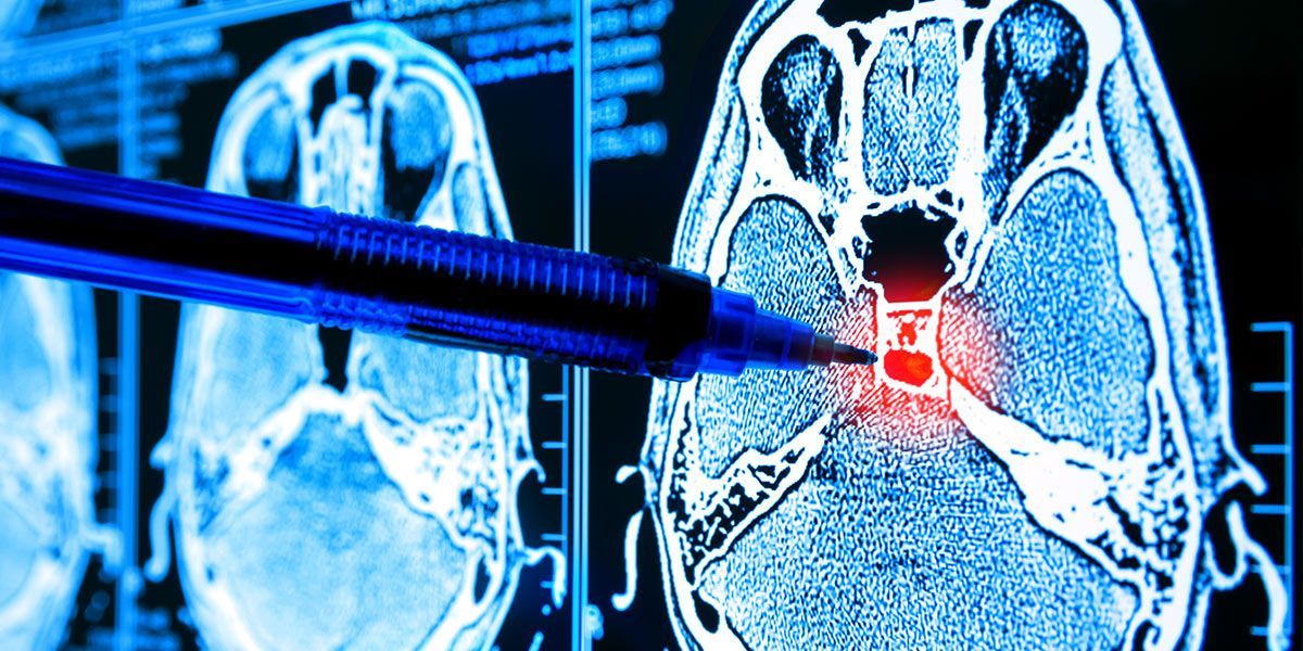
6. Brain Aneurysms
A traumatic brain injury, or TBI, is usually caused by a violent blow or penetration to the head and causes brain disarray. According to Medline Plus, some common causes for these severe head injuries include falls, sports injuries, child abuse, being struck by an object, or being hit or shot with a weapon.
A newer technique using contrast agents has helped physicians visualize the damage of a TBI or concussion. Essentially, a contrast agent is a dye that can be injected or consumed before the MRI scan that makes the images from the MRI clearer.
At Upright MRI of Deerfield, a contrast agent may be necessary in order to make a proper diagnosis. In our experience, 5 to 10 percent of patients require a contrast agent.
7. Carotid Artery Disease
Carotid artery disease is when fatty deposits, or plaque, builds up and clogs blood vessels that deliver blood to your brain and head, which eventually may lead to a stroke or heart attack.
The images the MRI scans produce allows doctors to visualize this blockage in blood vessels and determine treatments before the blockage becomes severe and life threatening.
8. Herniated Discs
IF you have herniated discs, this means there is a problem with the rubbery part of your spine between the vertebrae, causing irritation, pain or weakness in the arm or leg.
The specific cause is hard to determine, but it’s something that occurs slowly over time. It could be from having a physically demanding job where you are twisting your muscles and turning your spine everyday, or simply from aging. As we age, the disks (the rubbery part) become more flexible and therefore more susceptible to tearing.
One of the great benefits of having an Open Upright MRI is that it allows patients to be scanned in any position, therefore allowing pretty much any part of the body to be imaged. Because of this, patients can lie down comfortably or be scanned standing or sitting when they have a herniated disc.

9. Fractures
A fracture is a break in the bone, often caused by accidents or injuries, or diseases that weaken the bones.
An X-ray can diagnose bone fractures, but MRIs can provide a more detailed image of the bone, which helps physicians understand exactly where the break is and how to properly treat it.
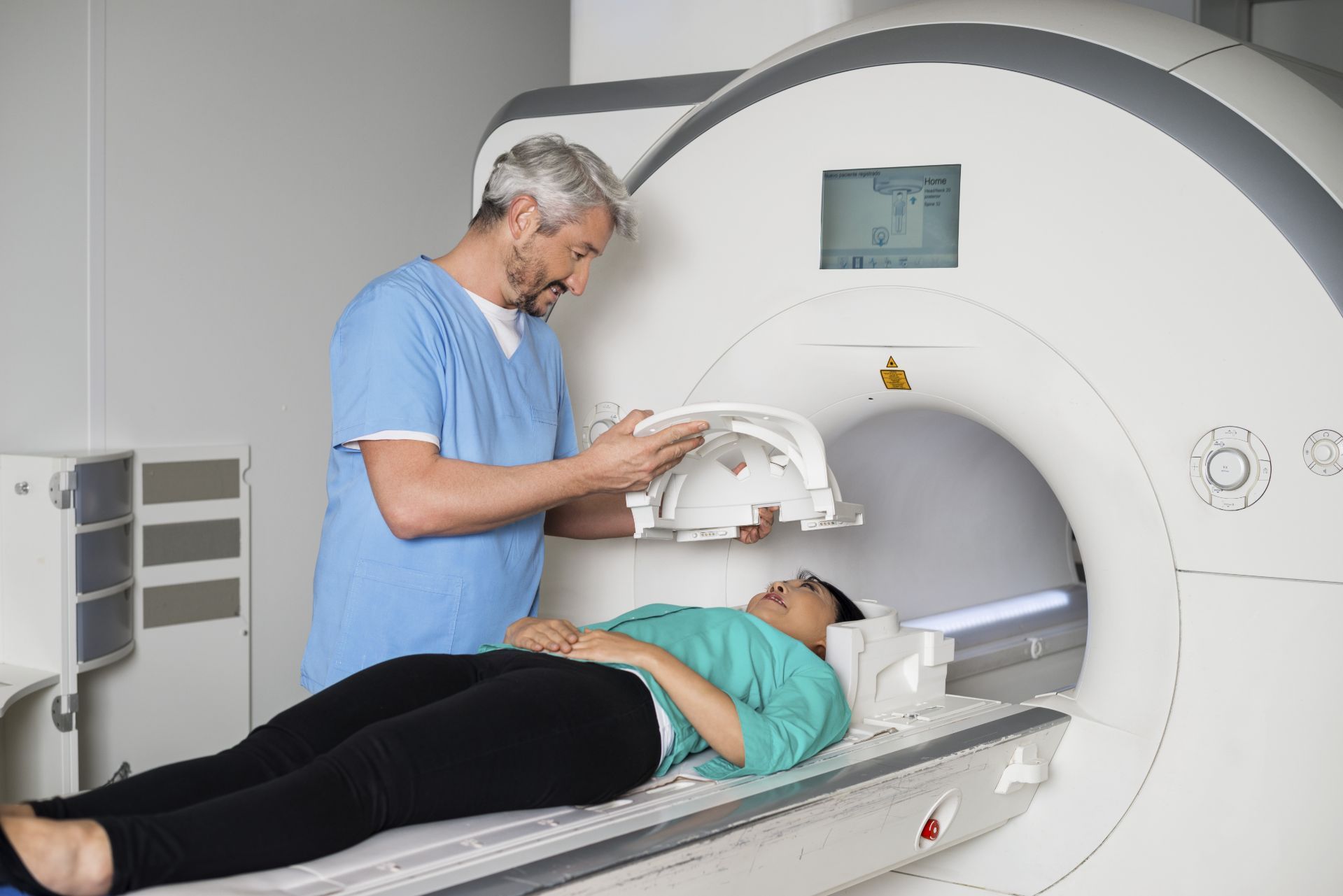
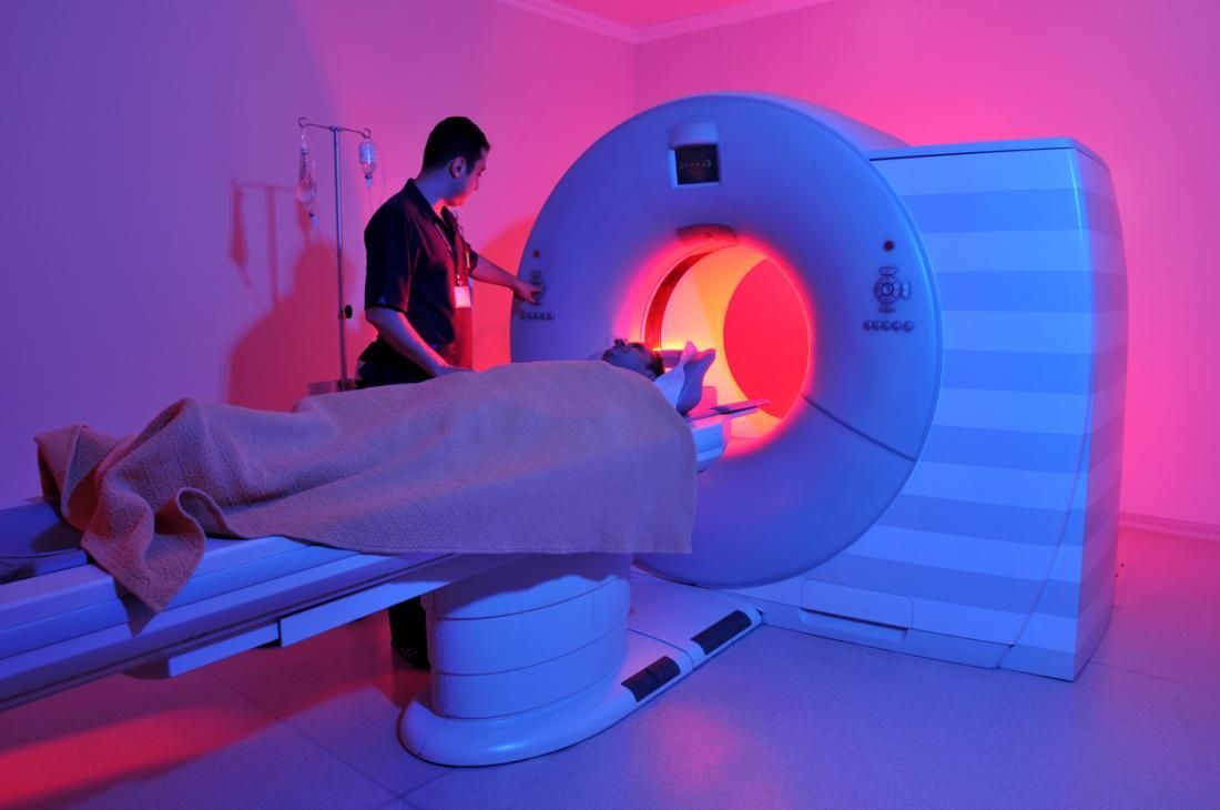
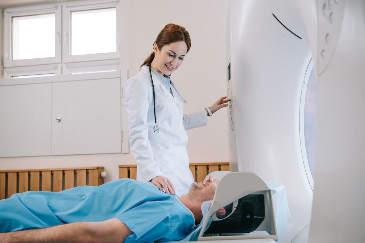
MRI is a key neuroimaging tool that allows doctors to get a solid understanding of what is happening in your body. At Upright MRI of Deerfield, we use the FONAR UPRIGHT® Multi-Position™ MRI, which allows the patient to comfortably sit or stand in several different positions.
This allows us to see what is happening from several different angles and makes the MRI experience much more relaxed and open for our patients.
To learn more about our MRI and schedule a visit, go to
www.uprightmrideerfield.com.

SHARE THIS POST:
Leave a Comment:
The World's Most Patient-Friendly MRI. A comfortable, stress-free, and completely reliable MRI scan. We offer patients an open, upright, standup MRI experience that helps those who are claustrophobic and stress being in a confined area. Upright MRI of Deerfield is recognized as the world leader in open MRI innovation,
Our Recent Post


READ PATIENT TESTIMONIALS
Upright MRI of Deerfield.
Susan D.,
Highland Park, 39
I am going to tell everyone about your office! This was a great experience after I panicked in other MRI machines and had to leave. Thank you so much.

Judith B.,
Milwaukee, 61
I suffer from vertigo and other MRIs do not work. This was wonderful…absolutely NO discomfort at all. The MRI was so fast…I wanted to stay and watch the movie! Mumtaz was great. His humor really put me at ease. I’ve already recommended Upright MRI to friends.
Delores P.,
Glencoe, 55
Everything is so nice and professional with your place. I have been there a couple of times. My husband and I would not go anywhere else.

Follow UpRight MRI of Deerfield on Facebook
To see our latest news, updates or to get to know us more, we welcome you to follow along our journey in Facebook.


CONTACT DETAILS
Phone: (847) 291-9321
Address: 457 Lake Cook Road (Deerfield Park Plaza) Deerfield, IL 60015
Email: info@uprightmrideerfield.com
Business Hours
- Mon - Thu
- -
- Friday
- -
- Saturday
- -
- Sunday
- Closed