
Written by Geunu Jeong
Head of Research in AIRS Medical
Taking a brain MRI at the hospital is not just about capturing a single image. Various images are taken with different purposes and uses (refer to Why does it take so long for an MRI scan?). These images are then comprehensively assessed to create a diagnostic report. The composition of brain MRI images varies depending on what needs to be observed. Typically, brain MRI includes T1-weighted images, T2-weighted images, FLAIR images, and contrast-enhanced T1-weighted images. The abundance of technical terms might seem overwhelming, but don’t worry. In this post, the focus is not on understanding the technical jargon but on explaining each image’s clinical purposes and uses for easy comprehension.
1. T1-weighted images
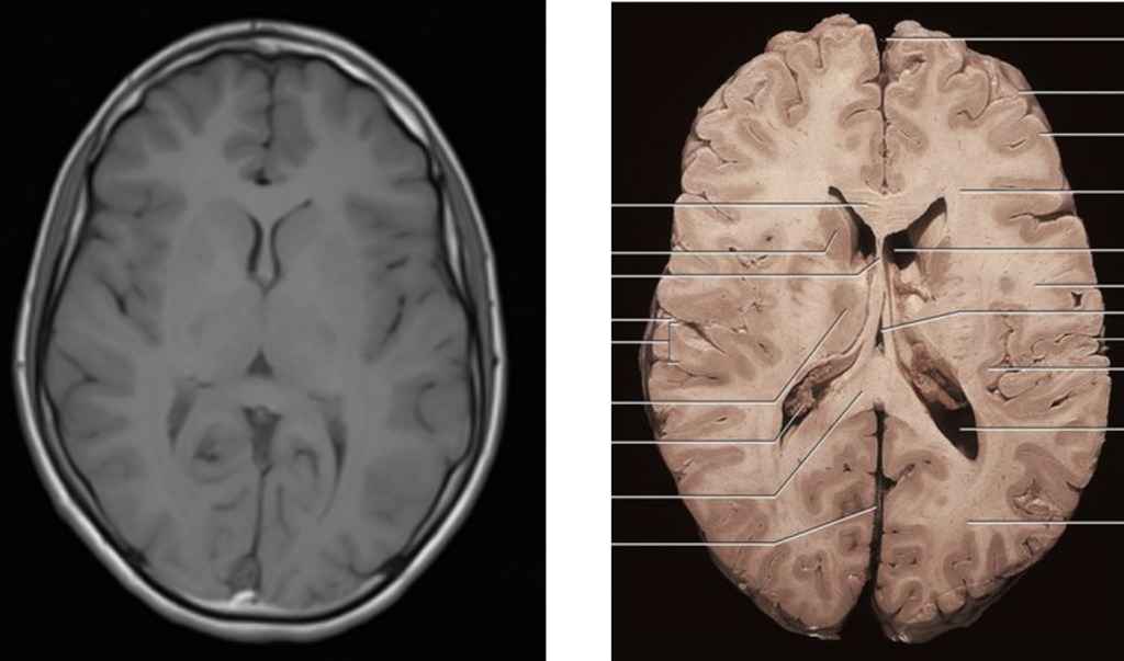
T1-weighted images provide the best representation of the actual anatomical structures. On the left is a T1-weighted MRI image, and on the right is an actual cross-sectional photo of the brain. The white areas in the right photo are also brightly represented in the T1-weighted image, and the darker areas in color are similarly represented in the T1-weighted image. This helps to understand the brain’s structure intuitively.
2. T2-weighted images
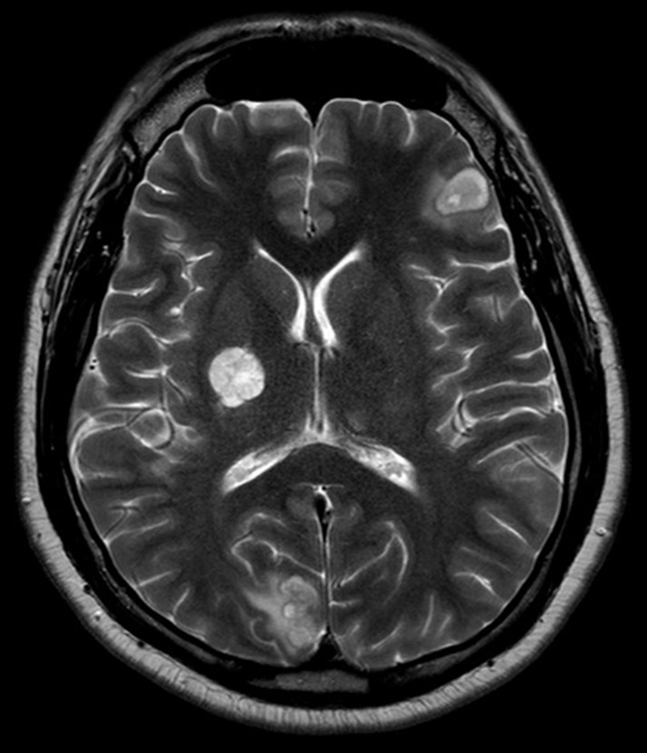
T2-weighted images are excellent for detecting lesions. Tissues with a higher water content appear bright in T2-weighted images, making it easier to identify most lesions, as they often have an increased water content in their early stages. In the above photo, you can also see a small lesion on the left side, represented as a coin-sized brightness area.
3. FLAIR images
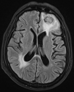
FLAIR (Fluid-Attenuated Inversion Recovery) images are essentially based on T2-weighted images but are designed to suppress cerebrospinal fluid (CSF), making it appear dark. In the central part of the image above are brain ventricles filled with a liquid like water, known as cerebrospinal fluid. In T2-weighted images, CSF appears bright because it is like water. However, in FLAIR images, it appears dark. The drawback of T2-weighted images is that CSF can appear too bright, making lesions relatively indistinct. In contrast, FLAIR images depict CSF as dark, making it easier to detect lesions.
4. Contrast-Enhanced T1-weighted images
In some cases, patients are injected with a contrast agent before imaging. However, contrast agent injection carries risks for the patient, so it is only done when the benefits outweigh the risks. This is primarily done when there is suspicion of tumors or inflammation.
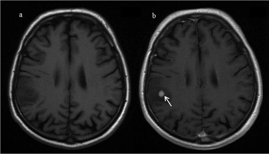
The left image is a T1-weighted image taken before administering the contrast agent, and the right image is a T1-weighted image taken after administration. The right image is referred to as a contrast-enhanced T1-weighted image. Detecting tumors in the left image is challenging, but they are easily identifiable in the right image. Moreover, contrast-enhanced images are useful for determining the type of tumor based on the presence and pattern of contrast enhancement.
How do you find it? This post briefly explored the purposes and uses of the different images that make up a brain MRI. They may all appear to be the same black-and-white images from a distance, but as you’ve seen up close, their purposes and functions are vastly different. I tried to explain it as simply as possible, but if you found it too difficult or have more questions, please let us know via email or contact us page. Any questions about MRI are always welcome!