Your doctor thinks it would be a good idea for you to get an MRI to make sure you don’t have a brain tumor.
We understand how intimidating and stressful this might be for you. Many people feel the same way when their doctor recommends a brain MRI.
Know that an MRI scan will give you and your doctor highly detailed information about your brain.
An MRI, or Magnetic Resonance Imaging, is a medical imaging technique that uses powerful magnets and radio waves to produce detailed images of the inside of the body.
MRI machines use a large magnet and radio waves to generate signals from atoms in the body. Then a computer attached to the MRI converts those signals into images that a healthcare professional can study and understand.
How can an MRI scan provide detailed images of the brain?
One of the remarkable aspects of an MRI scan is its incredible ability to generate extremely detailed images. This is particularly important when scanning complex structures like the brain.
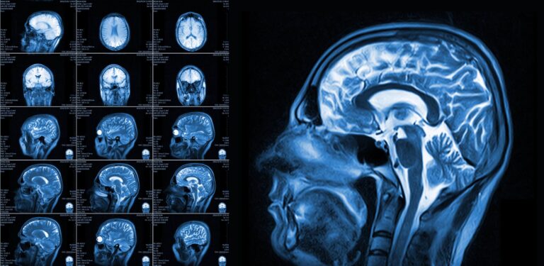
An MRI scan can differentiate between various types of tissues within the brain, thereby allowing doctors to identify any anomalies, such as tumors. It captures images in multiple planes, providing a comprehensive three-dimensional view of the brain.
MRIs are crucial for diagnosing brain tumors as they allow healthcare providers to locate the exact position, size, and extent of the potential tumor.
What are the benefits of using an MRI scan for brain tumor detection?
MRI scans offer several advantages for brain tumor detection. Their high-resolution, detailed images allow for accurate detection and characterization of tumors.
MRIs can even visualize soft tissues, blood vessels, and fluid buildup, providing comprehensive information about the condition of the brain. Because they do not use ionizing radiation, MRI scans are safer for patients than other imaging methods, particularly for those requiring multiple scans over time.
An MRI’s ability to scan in multiple planes helps doctors plan the best course of action for treatment or surgery, if necessary.
Are there any risks or limitations associated with MRI scans for brain tumors?
While MRI scans are generally considered safe and non-invasive, there are a few considerations and potential limitations.
Some individuals may feel uncomfortable or claustrophobic in the MRI machine due to the enclosed space and the noise it generates.
MRIs machines use a powerful magnet, so they cannot be used for patients with certain implants or devices like pacemakers or cochlear implants, unless these are marked as MRI-safe.
Although rare, people are sometimes allergic to contrast dye that some MRI procedures use. As with any medical procedure, it is important to discuss any potential risks or concerns with your healthcare provider.
Diagnosing brain tumors with an MRI
An MRI scan is an incredibly helpful tool for helping your doctor understand why you aren’t feeling well.
Let’s go over the facts of why doctors recommend brain MRIs, how a brain MRI can help show a possible tumor, and how MRIs can see tumors that could be concerning.
How does my doctor determine if I need an MRI scan for a possible brain tumor?
Several factors influence a doctor’s decision to recommend an MRI scan. These typically include the patient’s symptoms, medical history, and results of a physical examination.
If you’re experiencing symptoms such as persistent headaches, seizures, speech difficulties, loss of balance, vision problems, or other neurological symptoms, your doctor might suspect a brain tumor.
An MRI scan can help provide a detailed picture of your brain to better understand what might be causing these symptoms.
How can an MRI scan help identify a brain tumor?
An MRI scan offers a clear, detailed view of the brain’s tissues and structures. When a potential tumor is present, it appears as an abnormal growth differing from the surrounding brain tissue.
Additionally, a special dye called contrast material might be injected into your bloodstream during the MRI.
This dye travels to your brain and helps the tumor stand out in the images because the dye tends to accumulate more in the tumor cells than in the normal brain cells, highlighting any abnormal growths.
This is how your doctor figures out the location, size, and shape of the tumor.
How does an MRI differentiate between a malignant and a benign brain tumor?
MRI scans can provide important clues about a tumor’s nature—whether it’s benign (non-cancerous) or malignant (cancerous).
While an MRI cannot definitively diagnose cancer, it can help guide doctors.
Malignant tumors tend to grow rapidly and disrupt the normal functioning of surrounding brain tissues. They appear differently on MRI images compared to benign tumors, often showing as irregular, odd-looking shapes.
An MRI that uses contrast material can help make it even clearer, as malignant tumors often absorb more of the contrast dye.
However, a definitive diagnosis often requires additional tests, such as a biopsy, where a tiny sample of the tumor is studied under a microscope.
Can an MRI scan detect a brain tumor in the early stages?
Yes, an MRI scan can detect brain tumors in their early stages due to its high-resolution imaging capabilities.
The sensitivity of the MRI allows it to pick up small abnormalities, including tumors, much before they cause any noticeable symptoms.
However, detecting a brain tumor also depends on its location and type, and not all tumors may be detectable in their very early stages.
Keep in mind that routine scans are not usually recommended unless you’re at a high risk due to genetic predisposition or specific medical conditions.

Preparing for an MRI scan
On the day of your brain MRI, make sure you’re ready for your scan, to make sure everything goes quickly, and you get great results.
To get ready for your MRI, you’ll want to make sure that you’re dressed properly, that you’re following the right diet, and that your radiology technologist knows about your medical condition.
What should I do before my MRI scan?
First and foremost, you should wear comfortable and loose-fitting clothing, free of any metal zippers, buttons, or fastenings, as metal objects.
Anything made of metal can interfere with the MRI machine’s magnetic field. You may also be asked to remove jewelry, glasses, dentures, and hearing aids for the same reason.
Sometimes, the clinic may provide a hospital gown for you to wear during the scan.
Before your scan, try to minimize the use of hair products and skin products, as some may contain metallic particles.
Should I follow any dietary restrictions or precautions before the scan?
Unless specifically instructed by your healthcare provider, there are usually no dietary restrictions before an MRI scan.
However, if your scan involves the use of a contrast dye, you might be asked to fast for a few hours before the procedure. Always make sure to follow the instructions given by your doctor or the MRI center.
It’s also a good idea to stay well-hydrated, unless otherwise directed, and avoid caffeine if you’re prone to anxiety or restlessness.
Do I need to inform the medical staff about any pre-existing conditions or medications?
Yes, it’s absolutely vital to inform the medical staff about any pre-existing condition.
Tell a staff member if you have implants (for example, a pacemaker, cochlear implants, or vascular clips), metal fragments in your body, or any other metallic implants. The strong magnetic field of the MRI machine may interact with these devices.
It’s also important to inform the staff about any medications you’re currently taking, allergies, and whether you’re pregnant or suspect you may be.
If you have kidney disease, it’s particularly important to notify your healthcare team as this may affect your body’s ability to eliminate the contrast dye if one is to be used.
The MRI scan process
A brain MRI is nothing to stress over, but if you’ve never had one, then it’s easy to understand why you might be a little worried.
Let’s take a minute to get a closer look at what happens during a brain MRI, like what it’s like to get a brain scan, and how long your MRI will take.
What should I expect during my MRI scan?
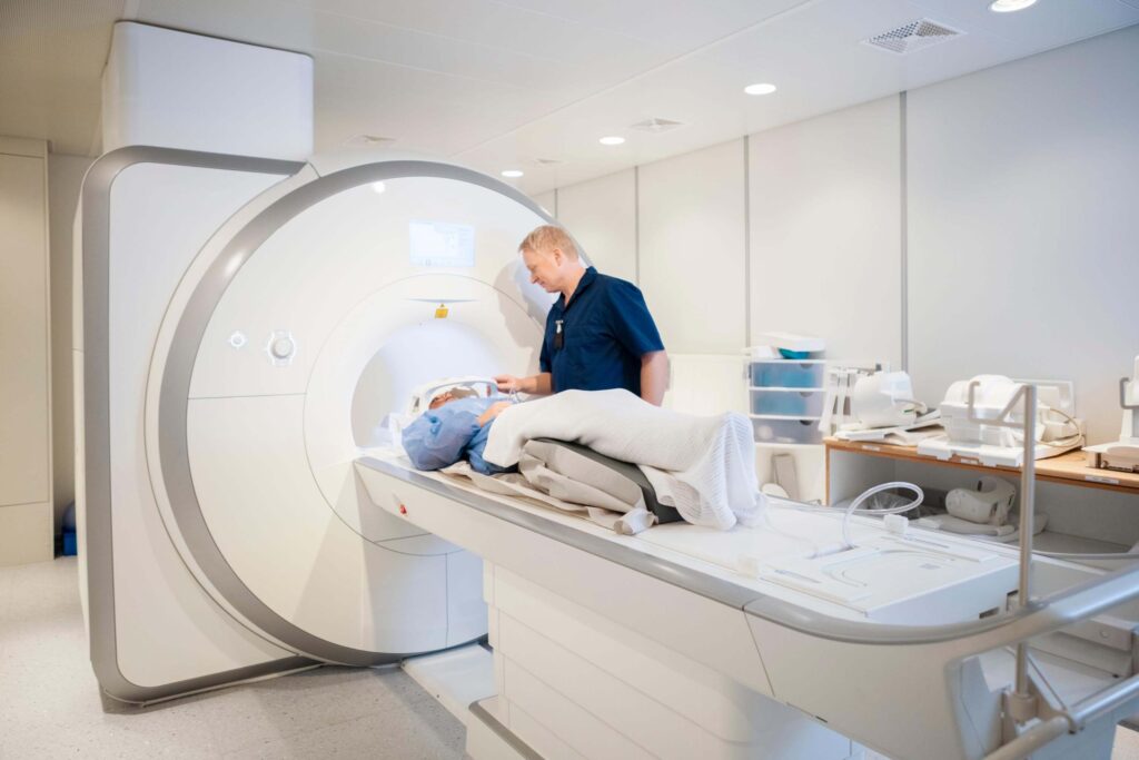
During the MRI scan, you’ll lie down on a motorized table that slides into a large, cylindrical machine. Before the scan starts, you might be given earplugs or headphones to minimize the noise produced by the machine.
If the scan requires a contrast dye, you may need to drink an oral contrast solution or have an intravenous (IV) line will be put into your arm to administer the dye during the scan.
Once you’re positioned and ready, the MRI technologist will leave the room but continue to communicate with you via an intercom. Throughout the scan, you’ll need to stay very still to ensure the images are clear and precise.
How do I need to position myself inside the MRI machine?
Your position in the MRI machine depends on the part of the body being scanned.
To scan for a potential brain tumor, you’ll be asked to lay on your back, with your head slightly elevated and supported by a padded holder.
You might also be fitted with a device known as a coil, which helps improve the quality of the images.
These are designed to be as comfortable as possible, but always communicate with your MRI technologist if you’re feeling uncomfortable.
Will the MRI scan be noisy or uncomfortable?
The MRI machine does produce a substantial amount of noise, often described as loud tapping or knocking sounds. This is why you’ll be provided with earplugs or headphones.
As for comfort, the inside of the MRI machine can feel a bit confined, and some people might experience feelings of claustrophobia.
If this is a concern, discuss it with your healthcare provider in advance. If you feel claustrophobic, try to relax yourself. In some cases, your healthcare provider may provide you with a mild sedative to take prior to your scan.
How long does the MRI scan typically take to complete?
The duration of an MRI scan can vary greatly depending on what is being imaged and whether a contrast material is used. Generally, for brain scans, you can expect the process to last between 12 to 20 minutes.
However, you should plan on being at the facility for around two hours to allow time for preparation, the scan itself, and any post-scan procedures.
Understanding your brain MRI results
After your scan, it will take a few days for your doctor to get your MRI results back from the imaging center.
During that time, your doctor will take a closer look at your results, and then contact you. Let’s look at how long the results will take, what your doctor is looking for, and what happens next.
How long will it take for me to receive my MRI results?
The turnaround time for MRI results can vary depending on factors like the complexity of the images, the need for further analyses, and the diagnostic center.
Typically, a radiologist will examine your MRI images within 24 to 48 hours, and your healthcare provider will usually receive the results within this timeframe.*
Once your doctor has the results, they will arrange a follow-up appointment to discuss the findings with you.
What is my doctor looking for in my MRI results?
MRI images can provide intricate details of the brain’s structure, making them valuable for diagnosing brain tumors. In the MRI images, a brain tumor typically appears as an abnormal mass or growth.
Your healthcare provider will look for any such irregularities, taking note of their size, shape, and location. They will also examine the MRI images to assess whether the tumor is affecting other structures within the brain.
Contrast-enhanced MRI scans are especially useful because they can help distinguish tumor tissue from surrounding healthy tissue, making it easier to identify and characterize potential brain tumors.
Will I need additional tests or consultations after my MRI scan?
The need for additional tests or consultations largely depends on your MRI results. If your doctor suspects a possible brain tumor, they may recommend further diagnostic tests to gather more information.
This might include a biopsy, where a small sample of the tumor is removed for analysis to determine the type and grade of the tumor.
In some cases, if the MRI results are inconclusive or if the potential tumor is in a challenging location, your provider may suggest additional imaging tests like a CT scan or a PET scan to get a more comprehensive view.
You might also be referred to a neurologist or a neurosurgeon for a more specialized consultation.
Support and follow-up after your brain MRI
Lots of people feel stress when it comes to getting a brain MRI.
No matter what the results are, it’s hard not to feel worried and overwhelmed, and that’s why we’re here to help you.
Let’s take a look at some simple ways you can deal with waiting for your results, at what you can do if your doctor has any concerns, and at how you can get a second opinion.
How can I cope with feeling anxious or stressed while I wait for my MRI results?
The waiting period for results can be a stressful time, but there are strategies you can employ to help manage your anxiety.
Breathing exercises and mindfulness techniques can be effective in the short term, helping to reduce immediate stress levels.
Regular exercise and maintaining a balanced diet can also be beneficial.
Connecting with loved ones or joining support groups where you can share your feelings and experiences with others going through a similar situation can be immensely comforting.
If your anxiety becomes overwhelming, please don’t hesitate to seek professional help from a counselor or psychologist who specializes in helping individuals coping with medical-related stress.
What are the possible next steps if my doctor detects a brain tumor on my MRI scan?
If a suspected brain tumor is detected on your MRI scan, your doctor will likely refer you to a specialist such as a neurologist or neurosurgeon.
They will work with you to develop a detailed treatment plan based on the type, size, and location of the tumor, as well as your overall health.
Treatment options may include surgery, radiation therapy, chemotherapy, or a combination of these.
Your healthcare team will explain all available options and potential side effects so you can make an informed decision about your treatment.
After a brain tumor diagnosis, is it OK to get a second opinion?
Absolutely, getting a second opinion is not only OK but often recommended.
Brain tumors and their treatment plans can be complex, and seeking a second opinion can provide reassurance, or offer alternative treatment options.
Doctors are familiar with this practice and should support your decision to seek additional advice.
Remember, you don’t have to navigate this journey alone. Reach out to healthcare professionals, connect with support groups, and ask for help from your loved ones.
How to Schedule a Brain MRI Appointment with Us
Reach out to us at American Health Imaging, and we’ll help you schedule an appointment at an imaging center near you, today.
Did you know our MRIs cost up to 60% less than what you pay at a hospital?
We’re here to help you get the answers you need.
Frequently Asked Questions
What exactly is an MRI scan and how does it function?
An MRI (Magnetic Resonance Imaging) is a type of scan that uses magnetic fields and radio waves to create detailed images of structures within the body, such as the brain.
What is it about an MRI scan that allows it to produce detailed images of the brain?
MRI scans are able to create detailed brain images due to their ability to detect variations in water content and distribution in various tissues and structures.
How does a doctor decide whether an MRI scan is necessary for diagnosing a brain tumor?
A doctor typically orders an MRI scan if a patient presents symptoms that may indicate a brain tumor, such as persistent headaches, seizures, or cognitive difficulties.
Can MRI scans differentiate between malignant and benign brain tumors?
Yes, MRI scans can often differentiate between malignant and benign tumors by analyzing the tumor’s shape, size, and the way it interacts with the surrounding tissue.
Are there any special instructions I should follow before my MRI scan?
Yes, you may need to fast for a certain period of time, avoid certain medications, and remove all metal objects from your body prior to the scan.
What should I expect during my MRI scan?
During an MRI scan, you will be asked to lie still inside the MRI machine, which is a large tube, and you may hear loud noises as the machine works.
If my doctor suspects a brain tumor, what will they be looking for in my MRI results?
A doctor will be looking for abnormalities such as unusual shapes, sizes, or growth patterns, which could indicate the presence of a brain tumor.
If my MRI scan indicates a brain tumor, what are the possible next steps?
If a brain tumor is detected, the next steps can include further diagnostic tests, consultations with specialists, and discussion of treatment options, which may include surgery, radiation therapy, or chemotherapy.