SlideShare a Scribd company logo
1 of 29
VETERINARY COLLEGE HASSAN
VETERINARYANATOMY
SUBMITTED TO:
Dr Rajashailesh N M
HOD
Dept of Veterinary Anatomy
SUBMITTED BY:
Swaroop R
Jr MVSc
Dept of VSR
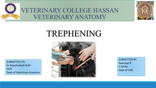
TREPHENING
Trephining is the surgical procedure in which a hole is created in the skull by the removal of
circular piece of bone, while a trepanation is the opening created by this procedure
(Stone and Miles, 1990).
TREPHENING
Trephination (also known as trepanning or burr holing) is a
surgical intervention where a hole is drilled, incised or scraped
into the skull using simple surgical tool
Sinuses: Anatomy
There are five pairs of paranasal sinuses :
1. Frontal
2.Maxillar
3.Sphenopalatine
4. Ethmoidal
5. Lacrimal
The topographical anatomy of the frontal and maxilary sinuses is of clinical importance.
The empyma of these sinuses is very common in the domestic animals.
Hence there is need to trephine open these sinuses to flush out the exudate.
Sinuses of cattle
1.Cornual diverticulum of frontal
sinus
2. Frontal sinus
3. Anterior-medial part of frontal
sinus
4. Lacrimal sinus
5. Maxillary sinus
6. Palatine sinus.
TREPHINATION OF PARANASAL SINUSES
The equine paranasal sinuses (PNS) are an intricate area of the head.
All of these spaces communicate with each other and the nasal passage either directly or
indirectly.
 A thorough understanding of the spatial structural relationships will improve one's ability to
interpret imaging studies and plan the best approach to the PNS.
 Trephination of the PNS is a basic procedure that can be readily performed in the field, and
familiarity with anatomy is the most important aspect of the procedure.
INDICATIONS
1. Sinusitis and pus in the sinuses.
2. Neoplasms in the sinuses.
3. Fracture of related bones.
4. Horn cancer in cattle and buffalo.
5. Dental fistula.
6. Alveolar periostitis.
7. Presence of any foreign body/cyst/parasite in sinuses.
SURGICALANATOMY-HORSE
 Frontal sinus in horse is not so extensive as that
of cattle.
The median septum divides right and left
sinuses.
The frontal sinus is divided into frontal and
turbinate parts and further sub divided in small
compartments by number of body plates.
Each compartment communicates with each
other by small openings.
The frontal bone lines the roof of sinus which
extends anteriorly with anterior margin of orbit
and caudally to temporal condyles and laterally
to the foot of the supraorbital process.
1.Frontal sinus
2. Anterior compartment of maxillary sinus;
3. Posterior compartment of maxillary sinus;
4. Palatine sinus.
Sinuses of horse.
 The turbinate part is located in the posterior part of the dorsal turbinate bone
covered by nasal and lacrimal bones.
 The turbinate part is separated from the nasal cavity by a thin tissue of dorsal
turbinate bone.
 The frontal and maxillary sinuses communicate with each other through a large
fronto-maxillary opening, which is situated ventral to the osseous canal and
medial wall of the orbit.
The frontal sinus has no direct communication with the nasal cavity.
Site for trephining
The point of frontal sinus trephine is placed 2.5cm lateral to the midline of skull on a horizontal
line running from the upper border of the orbit across to a similar point on the other side.
SURGICAL TECHNIQUE - FRONTAL
SINUS
1. Make a ‘V’ shaped skin incision at the site of trephining and expose the periosteum.
2. With the help of a trephine of appropriate size open the frontal sinus through the turbinate
portion.
3. Establish a communication between the sinus and nasal cavity with a curved mare catheter.
Apply slight pressure to break the median septum, felt with finger through the trephining opening.
4. Place a gauge in the sinus and daily irrigation with 1:1000 potassium permanganate may be
done till complete infection is removed.
5. Skin wound is sutured after complete removal of the infection
left; Under standing sedation following skin preparation
and local anaesthesia, a 3 cm vertical skin and periosteal
incision is made over the frontal sinus.
Right; a 10 mm diameter, modified tungsten carbide
metalwork drill bit used for sinus trephination.
Use of modified drill to enter the CFS. The welded flange,
approximately 3 cm from its tip, prevents the drill from
suddenly penetrating the sinus too deeply and possibly
damaging the ethmoturbinates.
Horse with sinus lavage
catheter inserted in its right
concho-frontal sinus.
Sinoscopy (using a frontal sinus portal) shows a single piece of
inspissated pus lying on the floor of the caudal maxillary sinus
of a horse with chronic low-grade primary sinusitis. The
sinusitis resolved following its removal using transendoscopic
biopsy forceps
SURGICALANATOMY FRONTAL SINUS
CATTLE AND BUFFALO
a) Largest and involves whole of frontal bone.
b) Median septum separates right & left sinuses.
c) The boundaries of frontal sinus are:
i. Anteriorly, an imaginary transverse line from inner canthus of one orbit to the other.
ii. Posteriorly squamous part of the occipital bone.
iii. Laterally, supraorbital process and the lateral border of frontal bone.
iv. Medially, median septum.
v. Caudally sinus extends for a variable distance into cornual process.
d) The sinus is very irregular and is divided into numerous diverticulae by ridges and
partial septae.
e) Several small openings from middle nasal meatus communicate directly with each
sinus.
f) The anterior portion of frontal sinus communicates with the lacrimal part of
maxillary sinus through a large opening which remains closed by a mucous membrane
in living state.
SITE OF OPERATION FRONTAL SINUS
Cattle & Buffalo
a) 4 cm from the posterior edge of the orbital cavity and just dorsal to the temporal canthus or
where the bulging of bone is seen to drain out postorbital diverticulum.
b) Posterior to a line joining the center of the orbit and 2-3 cm from the mid line to drain out
medial part.
c) For drainage of turbinate part-site is same as in horse.
1.Opening for main portion of frontal sinus
2.Frontal sinus
3. Opening for lowest portion of frontal sinus
4. Opening for anterior frontal sinus
5. Opening for turbinate portion of frontal sinus
6. Opening for maxillary sinus.
Location for trephine sites in the sinuses of cattle
MAXILLARY SINUS
a) Maxillary sinus is the largest sinus in horse. It is divided into anterior and posterior compartments by an
oblique septum.
b) It is formed by the superior maxillary, lacrimal, malar and posterior turbinate bones.
c) The boundaries of this sinus are:
i) Medially- Maxilla, ventral turbinate and lateral mass of the ethmoid bone.
ii) Posteriorly – The border extends upto transverse plane in front of root of the supraorbital process.
iii) Anteriorly- Line drawn from the anterior end of the facial crest to the infraorbital foramen parallel to the
facial crest.
iv) Ventrally- Alveolar part of maxilla.
d) The sinus is irregular and four cheek teeth project into it.
e) The sinus communicates with frontal sinus and nasal cavity.
SITE OF OPERATION
The anterior maxillary sinus is trephined at above 2.5-3.0 cm posterior and 2 cm medial to the
lower end of the zygomatic ridge.
b) Posterior maxillary sinus is trephined about 2 cm medial to the lower end of the zygomatic
ridge
Maxillary sinusotomy in a 3-year-old horse showing the small size
of both maxillary sinuses and the intervening maxillary septum.
The lateral aspect of the alveoli are approximately 1 cm from the
maxillary wall
Trephination of the left rostral maxillary sinus. Left: Skin and
muscle incision in an aseptically prepared site. Right: Drilling
through the maxillary bone using a soft tissue guard to reduce
skin and muscle damage
Inspissated pus being removed from a horse with chronic
primary sinusitis via a fronto-nasal sinusotomy
Standing sinusotomy of the rostral and caudal maxillary sinuses
in a horse with primary sinusitis has allowed mucoid and
mucopurulent exudate to drain from the caudal maxillary and
rostral maxillary sinuses, respectively
MAXILLARY SINUS - CATTLE
AND BUFFALO
a) It is situated anterior to orbital cavity and is formed by lacrimal, malar and the body of maxilla.
It is single and doesn’t communicate with frontal sinus.
b) It is irregular sinus. The roots of last three or four check teeth project into it.
c) The boundaries of maxillary sinus are: i) Dorsally – Lacrimal bulla, little below the point of
bifurcation of the zygomatic process of malar.
ii) Posteriorly- Base of alveoli and maxillary tuberosity.
iii) Anteriorly – Line joining infraorbital foramina to the orbital rim
. iv) Laterally- Maxilla, lacrimal and malar bones.
v) Medially- Irregular bone plates.
d) The maxillary and palatine sinuses communicate freely with each other
Trephination site for access to the bovine
maxillary sinus
 The landmarks for the trephination site for the bovine maxillary sinus are the medial canthus of the eye
and the facial tuberosity.
 A line is drawn between the facial tuberosity and the canthus.
 The trephination site is 1 cm ventral to the midway point of this line to avoid the nasolacrimal duct.
 The soft tissue and bone is considerably thicker than at the equivalent trephination sites (rostral and
caudal maxillary sinuses) in the horse
The left radiograph shows the continued presence of the fluid
line. The area of increased radioopacity seen previously is no
longer present. The trephination site is marked with a coin on
the right radiograph.
Lateral radiograph with the horizontal fluid line
(arrows) clearly visible in the right maxillary sinus.
Lateral radiograph taken after 10 days of antibiotic
therapy. The fluid line in the maxillary sinus is still
clearly visible. There appears to be an area of
increased radioopacity adjacent to the maxillary
sinus (arrows).
CONCLUSION
Anatomy of paranasal sinuses is essential for successful trephining.
Maxillary sinus is more affected in Horse
Frontal sinus more affected in Cattle
Post operative care is more essential
REFERENCES
Barakzai, S.Z., KANE‐SMYTH, J.U.S.T.I.N.E., Lowles, J. and Townsend, N., 2008. Trephination of the
equine rostral maxillary sinus: efficacy and safety of two trephine sites. Veterinary
Surgery, 37(3), pp.278-282.
Dixon, P.M. and O'Leary, J.M., 2012. A review of equine paranasal sinusitis: medical and surgical
treatments. Equine Veterinary Education, 24(3), pp.143-158.
Crilly, J.P. and Hopker, A., 2013. Case report: diagnosis and treatment of maxillary sinusitis in an
Aberdeen Angus cow. Livestock, 18(6), pp.230-235.
Applied Anatomy Of Domestic Animals-R L Bharadwaj, Rajesh Rajput,K S Roy
trephining.pptx

More Related Content

What's hot

Ear new affection of ear and its treatment
Ear new affection of ear and its treatmentEar new affection of ear and its treatment
Ear new affection of ear and its treatmentBikas Puri
 
Canine radiographs
Canine radiographsCanine radiographs
Canine radiographsblankita2010
 
Caessarean section in bovines
Caessarean section in bovinesCaessarean section in bovines
Caessarean section in bovinesIVRI
 
Dog vaginal cytology by Dr.mehdi moradi
Dog vaginal cytology by Dr.mehdi moradiDog vaginal cytology by Dr.mehdi moradi
Dog vaginal cytology by Dr.mehdi moradimehdi moradi
 
Traumatic reticuloperitonitis , traumatic pericarditis
Traumatic reticuloperitonitis , traumatic pericarditisTraumatic reticuloperitonitis , traumatic pericarditis
Traumatic reticuloperitonitis , traumatic pericarditisRekha Pathak
 
Cs small animals
Cs small animalsCs small animals
Cs small animalskgadipk88
 
Cystic ovarian degeneration Dr. Najmu Saaqib Reegoo DVM
Cystic ovarian degeneration  Dr. Najmu Saaqib Reegoo DVM Cystic ovarian degeneration  Dr. Najmu Saaqib Reegoo DVM
Cystic ovarian degeneration Dr. Najmu Saaqib Reegoo DVM Najamu Saaqib Reegoo
 
Lecture 24 Ultrasonography in animal reproduction
Lecture 24 Ultrasonography in animal reproductionLecture 24 Ultrasonography in animal reproduction
Lecture 24 Ultrasonography in animal reproductionDrGovindNarayanPuroh
 
lameness (equine hoof affections) Dr. Alaa Ghazy
lameness (equine hoof affections) Dr. Alaa Ghazylameness (equine hoof affections) Dr. Alaa Ghazy
lameness (equine hoof affections) Dr. Alaa GhazyDr. Alaa Ghazy
 
Caesarean section in cow
Caesarean section in cowCaesarean section in cow
Caesarean section in cowRekha Pathak
 
Equine med power point presentation
Equine med power point presentationEquine med power point presentation
Equine med power point presentationdevonavis
 
Intussusception in cattle
Intussusception in cattleIntussusception in cattle
Intussusception in cattleilyaszargar
 
Examination of superficial lymph nodes in dogs and cat
Examination of superficial lymph nodes in dogs and catExamination of superficial lymph nodes in dogs and cat
Examination of superficial lymph nodes in dogs and catGansbaai SA
 
Paravertebral nerve block in animals
Paravertebral nerve block in animalsParavertebral nerve block in animals
Paravertebral nerve block in animalsDR AMEER HAMZA
 
Diaphragmatic hernia
Diaphragmatic hernia Diaphragmatic hernia
Diaphragmatic hernia Rekha Pathak
 
4th Year - Theriogenology Practical Revision - 1st Term - Class 2013
4th Year - Theriogenology Practical Revision - 1st Term - Class 20134th Year - Theriogenology Practical Revision - 1st Term - Class 2013
4th Year - Theriogenology Practical Revision - 1st Term - Class 2013Mohamed Wahab
 

What's hot (20)

Ear new affection of ear and its treatment
Ear new affection of ear and its treatmentEar new affection of ear and its treatment
Ear new affection of ear and its treatment
 
Canine radiographs
Canine radiographsCanine radiographs
Canine radiographs
 
Caessarean section in bovines
Caessarean section in bovinesCaessarean section in bovines
Caessarean section in bovines
 
Colic in horses
Colic in horsesColic in horses
Colic in horses
 
Dog vaginal cytology by Dr.mehdi moradi
Dog vaginal cytology by Dr.mehdi moradiDog vaginal cytology by Dr.mehdi moradi
Dog vaginal cytology by Dr.mehdi moradi
 
Traumatic reticuloperitonitis , traumatic pericarditis
Traumatic reticuloperitonitis , traumatic pericarditisTraumatic reticuloperitonitis , traumatic pericarditis
Traumatic reticuloperitonitis , traumatic pericarditis
 
G Equine Restraint
G  Equine RestraintG  Equine Restraint
G Equine Restraint
 
Cs small animals
Cs small animalsCs small animals
Cs small animals
 
Cystic ovarian degeneration Dr. Najmu Saaqib Reegoo DVM
Cystic ovarian degeneration  Dr. Najmu Saaqib Reegoo DVM Cystic ovarian degeneration  Dr. Najmu Saaqib Reegoo DVM
Cystic ovarian degeneration Dr. Najmu Saaqib Reegoo DVM
 
Lecture 24 Ultrasonography in animal reproduction
Lecture 24 Ultrasonography in animal reproductionLecture 24 Ultrasonography in animal reproduction
Lecture 24 Ultrasonography in animal reproduction
 
Estrus cycle of bitch
Estrus cycle of bitchEstrus cycle of bitch
Estrus cycle of bitch
 
lameness (equine hoof affections) Dr. Alaa Ghazy
lameness (equine hoof affections) Dr. Alaa Ghazylameness (equine hoof affections) Dr. Alaa Ghazy
lameness (equine hoof affections) Dr. Alaa Ghazy
 
Caesarean section in cow
Caesarean section in cowCaesarean section in cow
Caesarean section in cow
 
Equine med power point presentation
Equine med power point presentationEquine med power point presentation
Equine med power point presentation
 
Intussusception in cattle
Intussusception in cattleIntussusception in cattle
Intussusception in cattle
 
Examination of superficial lymph nodes in dogs and cat
Examination of superficial lymph nodes in dogs and catExamination of superficial lymph nodes in dogs and cat
Examination of superficial lymph nodes in dogs and cat
 
Paravertebral nerve block in animals
Paravertebral nerve block in animalsParavertebral nerve block in animals
Paravertebral nerve block in animals
 
Diaphragmatic hernia
Diaphragmatic hernia Diaphragmatic hernia
Diaphragmatic hernia
 
Deg nala disease
Deg nala diseaseDeg nala disease
Deg nala disease
 
4th Year - Theriogenology Practical Revision - 1st Term - Class 2013
4th Year - Theriogenology Practical Revision - 1st Term - Class 20134th Year - Theriogenology Practical Revision - 1st Term - Class 2013
4th Year - Theriogenology Practical Revision - 1st Term - Class 2013
 

Similar to trephining.pptx

Ear discharge and otalgia
Ear discharge and otalgiaEar discharge and otalgia
Ear discharge and otalgiaDennis Lee
 
Anatomy of the external and middle ear
Anatomy of the external and middle earAnatomy of the external and middle ear
Anatomy of the external and middle earSalman Syed
 
Surgical anatomy of nose
Surgical anatomy of noseSurgical anatomy of nose
Surgical anatomy of noseAugustine raj
 
Maxillary sinus presentation
Maxillary sinus presentationMaxillary sinus presentation
Maxillary sinus presentationsiddharth verma
 
ANATOMY OF EXTERNAL AUDITORY CANAL
ANATOMY OF EXTERNAL AUDITORY CANALANATOMY OF EXTERNAL AUDITORY CANAL
ANATOMY OF EXTERNAL AUDITORY CANALKanu Saha
 
anatomyofnoseandparanasalsinuses-copy-230629065517-07a23e0b.pdf
anatomyofnoseandparanasalsinuses-copy-230629065517-07a23e0b.pdfanatomyofnoseandparanasalsinuses-copy-230629065517-07a23e0b.pdf
anatomyofnoseandparanasalsinuses-copy-230629065517-07a23e0b.pdfAliyahJohanis1
 
Anatomy of Nose And Paranasal Sinuses - Copy.pptx
Anatomy of Nose And Paranasal Sinuses - Copy.pptxAnatomy of Nose And Paranasal Sinuses - Copy.pptx
Anatomy of Nose And Paranasal Sinuses - Copy.pptxHtet Ko
 
Nasal cavity and paranasal sinuses
Nasal cavity and paranasal sinusesNasal cavity and paranasal sinuses
Nasal cavity and paranasal sinusesDentist Khawla
 
Nasal cavity and paranasal sinuses
Nasal cavity and paranasal sinusesNasal cavity and paranasal sinuses
Nasal cavity and paranasal sinusesDentist Khawla
 
Anatomy of nose (Applied)
Anatomy of nose (Applied)Anatomy of nose (Applied)
Anatomy of nose (Applied)BASIT ALI KHAN
 
Maxillary sinus
Maxillary sinusMaxillary sinus
Maxillary sinussauvik2014
 
Anatomy of Internal Nose
Anatomy of Internal Nose  Anatomy of Internal Nose
Anatomy of Internal Nose Arjun Graison
 
Maxillary sinus & its dental implication
Maxillary sinus & its dental implicationMaxillary sinus & its dental implication
Maxillary sinus & its dental implicationFiras Kassab
 
Anatomy Of Nose And Paranasal Sinuses - Copy.pptx
Anatomy Of Nose And Paranasal Sinuses - Copy.pptxAnatomy Of Nose And Paranasal Sinuses - Copy.pptx
Anatomy Of Nose And Paranasal Sinuses - Copy.pptxHtet Ko
 
Anatomy of Nose & Paranasal sinuses.pptx
Anatomy of Nose & Paranasal sinuses.pptxAnatomy of Nose & Paranasal sinuses.pptx
Anatomy of Nose & Paranasal sinuses.pptxHtet Ko
 

Similar to trephining.pptx (20)

Ear discharge and otalgia
Ear discharge and otalgiaEar discharge and otalgia
Ear discharge and otalgia
 
osteology.pptx
osteology.pptxosteology.pptx
osteology.pptx
 
Anatomy of the external and middle ear
Anatomy of the external and middle earAnatomy of the external and middle ear
Anatomy of the external and middle ear
 
Surgical anatomy of nose
Surgical anatomy of noseSurgical anatomy of nose
Surgical anatomy of nose
 
Imaging in ent
Imaging in entImaging in ent
Imaging in ent
 
Maxillary sinus
Maxillary sinusMaxillary sinus
Maxillary sinus
 
External rhinoplasty
External rhinoplastyExternal rhinoplasty
External rhinoplasty
 
Maxillary sinus presentation
Maxillary sinus presentationMaxillary sinus presentation
Maxillary sinus presentation
 
ANATOMY OF EXTERNAL AUDITORY CANAL
ANATOMY OF EXTERNAL AUDITORY CANALANATOMY OF EXTERNAL AUDITORY CANAL
ANATOMY OF EXTERNAL AUDITORY CANAL
 
Maxillary sinus sinus
Maxillary sinus sinus Maxillary sinus sinus
Maxillary sinus sinus
 
anatomyofnoseandparanasalsinuses-copy-230629065517-07a23e0b.pdf
anatomyofnoseandparanasalsinuses-copy-230629065517-07a23e0b.pdfanatomyofnoseandparanasalsinuses-copy-230629065517-07a23e0b.pdf
anatomyofnoseandparanasalsinuses-copy-230629065517-07a23e0b.pdf
 
Anatomy of Nose And Paranasal Sinuses - Copy.pptx
Anatomy of Nose And Paranasal Sinuses - Copy.pptxAnatomy of Nose And Paranasal Sinuses - Copy.pptx
Anatomy of Nose And Paranasal Sinuses - Copy.pptx
 
Nasal cavity and paranasal sinuses
Nasal cavity and paranasal sinusesNasal cavity and paranasal sinuses
Nasal cavity and paranasal sinuses
 
Nasal cavity and paranasal sinuses
Nasal cavity and paranasal sinusesNasal cavity and paranasal sinuses
Nasal cavity and paranasal sinuses
 
Anatomy of nose (Applied)
Anatomy of nose (Applied)Anatomy of nose (Applied)
Anatomy of nose (Applied)
 
Maxillary sinus
Maxillary sinusMaxillary sinus
Maxillary sinus
 
Anatomy of Internal Nose
Anatomy of Internal Nose  Anatomy of Internal Nose
Anatomy of Internal Nose
 
Maxillary sinus & its dental implication
Maxillary sinus & its dental implicationMaxillary sinus & its dental implication
Maxillary sinus & its dental implication
 
Anatomy Of Nose And Paranasal Sinuses - Copy.pptx
Anatomy Of Nose And Paranasal Sinuses - Copy.pptxAnatomy Of Nose And Paranasal Sinuses - Copy.pptx
Anatomy Of Nose And Paranasal Sinuses - Copy.pptx
 
Anatomy of Nose & Paranasal sinuses.pptx
Anatomy of Nose & Paranasal sinuses.pptxAnatomy of Nose & Paranasal sinuses.pptx
Anatomy of Nose & Paranasal sinuses.pptx
 

More from SwaroopR16

biomaterials in veterinary
biomaterials in veterinarybiomaterials in veterinary
biomaterials in veterinarySwaroopR16
 
humerus anatomy of domestic animals
humerus anatomy of domestic animalshumerus anatomy of domestic animals
humerus anatomy of domestic animalsSwaroopR16
 
OS COXAE.pptx
OS COXAE.pptxOS COXAE.pptx
OS COXAE.pptxSwaroopR16
 
BIOMECHANICS [Autosaved].pptx
BIOMECHANICS [Autosaved].pptxBIOMECHANICS [Autosaved].pptx
BIOMECHANICS [Autosaved].pptxSwaroopR16
 
computed tomography.pptx
computed tomography.pptxcomputed tomography.pptx
computed tomography.pptxSwaroopR16
 

More from SwaroopR16 (7)

pan.pptx
pan.pptxpan.pptx
pan.pptx
 
RUMENN.pptx
RUMENN.pptxRUMENN.pptx
RUMENN.pptx
 
biomaterials in veterinary
biomaterials in veterinarybiomaterials in veterinary
biomaterials in veterinary
 
humerus anatomy of domestic animals
humerus anatomy of domestic animalshumerus anatomy of domestic animals
humerus anatomy of domestic animals
 
OS COXAE.pptx
OS COXAE.pptxOS COXAE.pptx
OS COXAE.pptx
 
BIOMECHANICS [Autosaved].pptx
BIOMECHANICS [Autosaved].pptxBIOMECHANICS [Autosaved].pptx
BIOMECHANICS [Autosaved].pptx
 
computed tomography.pptx
computed tomography.pptxcomputed tomography.pptx
computed tomography.pptx
 

Recently uploaded

Grant Readiness 101 TechSoup and Remy Consulting
Grant Readiness 101 TechSoup and Remy ConsultingGrant Readiness 101 TechSoup and Remy Consulting
Grant Readiness 101 TechSoup and Remy ConsultingTechSoup
 
1029-Danh muc Sach Giao Khoa khoi 6.pdf
1029-Danh muc Sach Giao Khoa khoi  6.pdf1029-Danh muc Sach Giao Khoa khoi  6.pdf
1029-Danh muc Sach Giao Khoa khoi 6.pdfQucHHunhnh
 
Accessible design: Minimum effort, maximum impact
Accessible design: Minimum effort, maximum impactAccessible design: Minimum effort, maximum impact
Accessible design: Minimum effort, maximum impactdawncurless
 
Q4-W6-Restating Informational Text Grade 3
Q4-W6-Restating Informational Text Grade 3Q4-W6-Restating Informational Text Grade 3
Q4-W6-Restating Informational Text Grade 3JemimahLaneBuaron
 
Paris 2024 Olympic Geographies - an activity
Paris 2024 Olympic Geographies - an activityParis 2024 Olympic Geographies - an activity
Paris 2024 Olympic Geographies - an activityGeoBlogs
 
BAG TECHNIQUE Bag technique-a tool making use of public health bag through wh...
BAG TECHNIQUE Bag technique-a tool making use of public health bag through wh...BAG TECHNIQUE Bag technique-a tool making use of public health bag through wh...
BAG TECHNIQUE Bag technique-a tool making use of public health bag through wh...Sapna Thakur
 
Presentation by Andreas Schleicher Tackling the School Absenteeism Crisis 30 ...
Presentation by Andreas Schleicher Tackling the School Absenteeism Crisis 30 ...Presentation by Andreas Schleicher Tackling the School Absenteeism Crisis 30 ...
Presentation by Andreas Schleicher Tackling the School Absenteeism Crisis 30 ...EduSkills OECD
 
Sports & Fitness Value Added Course FY..
Sports & Fitness Value Added Course FY..Sports & Fitness Value Added Course FY..
Sports & Fitness Value Added Course FY..Disha Kariya
 
Nutritional Needs Presentation - HLTH 104
Nutritional Needs Presentation - HLTH 104Nutritional Needs Presentation - HLTH 104
Nutritional Needs Presentation - HLTH 104misteraugie
 
Introduction to Nonprofit Accounting: The Basics
Introduction to Nonprofit Accounting: The BasicsIntroduction to Nonprofit Accounting: The Basics
Introduction to Nonprofit Accounting: The BasicsTechSoup
 
Measures of Central Tendency: Mean, Median and Mode
Measures of Central Tendency: Mean, Median and ModeMeasures of Central Tendency: Mean, Median and Mode
Measures of Central Tendency: Mean, Median and ModeThiyagu K
 
1029 - Danh muc Sach Giao Khoa 10 . pdf
1029 -  Danh muc Sach Giao Khoa 10 . pdf1029 -  Danh muc Sach Giao Khoa 10 . pdf
1029 - Danh muc Sach Giao Khoa 10 . pdfQucHHunhnh
 
Sanyam Choudhary Chemistry practical.pdf
Sanyam Choudhary Chemistry practical.pdfSanyam Choudhary Chemistry practical.pdf
Sanyam Choudhary Chemistry practical.pdfsanyamsingh5019
 
Student login on Anyboli platform.helpin
Student login on Anyboli platform.helpinStudent login on Anyboli platform.helpin
Student login on Anyboli platform.helpinRaunakKeshri1
 
microwave assisted reaction. General introduction
microwave assisted reaction. General introductionmicrowave assisted reaction. General introduction
microwave assisted reaction. General introductionMaksud Ahmed
 
Measures of Dispersion and Variability: Range, QD, AD and SD
Measures of Dispersion and Variability: Range, QD, AD and SDMeasures of Dispersion and Variability: Range, QD, AD and SD
Measures of Dispersion and Variability: Range, QD, AD and SDThiyagu K
 
Web & Social Media Analytics Previous Year Question Paper.pdf
Web & Social Media Analytics Previous Year Question Paper.pdfWeb & Social Media Analytics Previous Year Question Paper.pdf
Web & Social Media Analytics Previous Year Question Paper.pdfJayanti Pande
 
The byproduct of sericulture in different industries.pptx
The byproduct of sericulture in different industries.pptxThe byproduct of sericulture in different industries.pptx
The byproduct of sericulture in different industries.pptxShobhayan Kirtania
 

Recently uploaded (20)

Grant Readiness 101 TechSoup and Remy Consulting
Grant Readiness 101 TechSoup and Remy ConsultingGrant Readiness 101 TechSoup and Remy Consulting
Grant Readiness 101 TechSoup and Remy Consulting
 
1029-Danh muc Sach Giao Khoa khoi 6.pdf
1029-Danh muc Sach Giao Khoa khoi  6.pdf1029-Danh muc Sach Giao Khoa khoi  6.pdf
1029-Danh muc Sach Giao Khoa khoi 6.pdf
 
Accessible design: Minimum effort, maximum impact
Accessible design: Minimum effort, maximum impactAccessible design: Minimum effort, maximum impact
Accessible design: Minimum effort, maximum impact
 
Q4-W6-Restating Informational Text Grade 3
Q4-W6-Restating Informational Text Grade 3Q4-W6-Restating Informational Text Grade 3
Q4-W6-Restating Informational Text Grade 3
 
Paris 2024 Olympic Geographies - an activity
Paris 2024 Olympic Geographies - an activityParis 2024 Olympic Geographies - an activity
Paris 2024 Olympic Geographies - an activity
 
BAG TECHNIQUE Bag technique-a tool making use of public health bag through wh...
BAG TECHNIQUE Bag technique-a tool making use of public health bag through wh...BAG TECHNIQUE Bag technique-a tool making use of public health bag through wh...
BAG TECHNIQUE Bag technique-a tool making use of public health bag through wh...
 
Presentation by Andreas Schleicher Tackling the School Absenteeism Crisis 30 ...
Presentation by Andreas Schleicher Tackling the School Absenteeism Crisis 30 ...Presentation by Andreas Schleicher Tackling the School Absenteeism Crisis 30 ...
Presentation by Andreas Schleicher Tackling the School Absenteeism Crisis 30 ...
 
Sports & Fitness Value Added Course FY..
Sports & Fitness Value Added Course FY..Sports & Fitness Value Added Course FY..
Sports & Fitness Value Added Course FY..
 
INDIA QUIZ 2024 RLAC DELHI UNIVERSITY.pptx
INDIA QUIZ 2024 RLAC DELHI UNIVERSITY.pptxINDIA QUIZ 2024 RLAC DELHI UNIVERSITY.pptx
INDIA QUIZ 2024 RLAC DELHI UNIVERSITY.pptx
 
Nutritional Needs Presentation - HLTH 104
Nutritional Needs Presentation - HLTH 104Nutritional Needs Presentation - HLTH 104
Nutritional Needs Presentation - HLTH 104
 
Introduction to Nonprofit Accounting: The Basics
Introduction to Nonprofit Accounting: The BasicsIntroduction to Nonprofit Accounting: The Basics
Introduction to Nonprofit Accounting: The Basics
 
Measures of Central Tendency: Mean, Median and Mode
Measures of Central Tendency: Mean, Median and ModeMeasures of Central Tendency: Mean, Median and Mode
Measures of Central Tendency: Mean, Median and Mode
 
1029 - Danh muc Sach Giao Khoa 10 . pdf
1029 -  Danh muc Sach Giao Khoa 10 . pdf1029 -  Danh muc Sach Giao Khoa 10 . pdf
1029 - Danh muc Sach Giao Khoa 10 . pdf
 
Sanyam Choudhary Chemistry practical.pdf
Sanyam Choudhary Chemistry practical.pdfSanyam Choudhary Chemistry practical.pdf
Sanyam Choudhary Chemistry practical.pdf
 
Student login on Anyboli platform.helpin
Student login on Anyboli platform.helpinStudent login on Anyboli platform.helpin
Student login on Anyboli platform.helpin
 
microwave assisted reaction. General introduction
microwave assisted reaction. General introductionmicrowave assisted reaction. General introduction
microwave assisted reaction. General introduction
 
Mattingly "AI & Prompt Design: Structured Data, Assistants, & RAG"
Mattingly "AI & Prompt Design: Structured Data, Assistants, & RAG"Mattingly "AI & Prompt Design: Structured Data, Assistants, & RAG"
Mattingly "AI & Prompt Design: Structured Data, Assistants, & RAG"
 
Measures of Dispersion and Variability: Range, QD, AD and SD
Measures of Dispersion and Variability: Range, QD, AD and SDMeasures of Dispersion and Variability: Range, QD, AD and SD
Measures of Dispersion and Variability: Range, QD, AD and SD
 
Web & Social Media Analytics Previous Year Question Paper.pdf
Web & Social Media Analytics Previous Year Question Paper.pdfWeb & Social Media Analytics Previous Year Question Paper.pdf
Web & Social Media Analytics Previous Year Question Paper.pdf
 
The byproduct of sericulture in different industries.pptx
The byproduct of sericulture in different industries.pptxThe byproduct of sericulture in different industries.pptx
The byproduct of sericulture in different industries.pptx
 

trephining.pptx

  • 1. VETERINARY COLLEGE HASSAN VETERINARYANATOMY SUBMITTED TO: Dr Rajashailesh N M HOD Dept of Veterinary Anatomy SUBMITTED BY: Swaroop R Jr MVSc Dept of VSR TREPHENING
  • 2. Trephining is the surgical procedure in which a hole is created in the skull by the removal of circular piece of bone, while a trepanation is the opening created by this procedure (Stone and Miles, 1990). TREPHENING Trephination (also known as trepanning or burr holing) is a surgical intervention where a hole is drilled, incised or scraped into the skull using simple surgical tool
  • 3. Sinuses: Anatomy There are five pairs of paranasal sinuses : 1. Frontal 2.Maxillar 3.Sphenopalatine 4. Ethmoidal 5. Lacrimal The topographical anatomy of the frontal and maxilary sinuses is of clinical importance. The empyma of these sinuses is very common in the domestic animals. Hence there is need to trephine open these sinuses to flush out the exudate.
  • 4. Sinuses of cattle 1.Cornual diverticulum of frontal sinus 2. Frontal sinus 3. Anterior-medial part of frontal sinus 4. Lacrimal sinus 5. Maxillary sinus 6. Palatine sinus.
  • 5. TREPHINATION OF PARANASAL SINUSES The equine paranasal sinuses (PNS) are an intricate area of the head. All of these spaces communicate with each other and the nasal passage either directly or indirectly.  A thorough understanding of the spatial structural relationships will improve one's ability to interpret imaging studies and plan the best approach to the PNS.  Trephination of the PNS is a basic procedure that can be readily performed in the field, and familiarity with anatomy is the most important aspect of the procedure.
  • 6. INDICATIONS 1. Sinusitis and pus in the sinuses. 2. Neoplasms in the sinuses. 3. Fracture of related bones. 4. Horn cancer in cattle and buffalo. 5. Dental fistula. 6. Alveolar periostitis. 7. Presence of any foreign body/cyst/parasite in sinuses.
  • 7. SURGICALANATOMY-HORSE  Frontal sinus in horse is not so extensive as that of cattle. The median septum divides right and left sinuses. The frontal sinus is divided into frontal and turbinate parts and further sub divided in small compartments by number of body plates. Each compartment communicates with each other by small openings. The frontal bone lines the roof of sinus which extends anteriorly with anterior margin of orbit and caudally to temporal condyles and laterally to the foot of the supraorbital process.
  • 8. 1.Frontal sinus 2. Anterior compartment of maxillary sinus; 3. Posterior compartment of maxillary sinus; 4. Palatine sinus. Sinuses of horse.
  • 9.  The turbinate part is located in the posterior part of the dorsal turbinate bone covered by nasal and lacrimal bones.  The turbinate part is separated from the nasal cavity by a thin tissue of dorsal turbinate bone.  The frontal and maxillary sinuses communicate with each other through a large fronto-maxillary opening, which is situated ventral to the osseous canal and medial wall of the orbit. The frontal sinus has no direct communication with the nasal cavity.
  • 10.
  • 11. Site for trephining The point of frontal sinus trephine is placed 2.5cm lateral to the midline of skull on a horizontal line running from the upper border of the orbit across to a similar point on the other side.
  • 12. SURGICAL TECHNIQUE - FRONTAL SINUS 1. Make a ‘V’ shaped skin incision at the site of trephining and expose the periosteum. 2. With the help of a trephine of appropriate size open the frontal sinus through the turbinate portion. 3. Establish a communication between the sinus and nasal cavity with a curved mare catheter. Apply slight pressure to break the median septum, felt with finger through the trephining opening. 4. Place a gauge in the sinus and daily irrigation with 1:1000 potassium permanganate may be done till complete infection is removed. 5. Skin wound is sutured after complete removal of the infection
  • 13. left; Under standing sedation following skin preparation and local anaesthesia, a 3 cm vertical skin and periosteal incision is made over the frontal sinus. Right; a 10 mm diameter, modified tungsten carbide metalwork drill bit used for sinus trephination. Use of modified drill to enter the CFS. The welded flange, approximately 3 cm from its tip, prevents the drill from suddenly penetrating the sinus too deeply and possibly damaging the ethmoturbinates.
  • 14. Horse with sinus lavage catheter inserted in its right concho-frontal sinus. Sinoscopy (using a frontal sinus portal) shows a single piece of inspissated pus lying on the floor of the caudal maxillary sinus of a horse with chronic low-grade primary sinusitis. The sinusitis resolved following its removal using transendoscopic biopsy forceps
  • 15. SURGICALANATOMY FRONTAL SINUS CATTLE AND BUFFALO a) Largest and involves whole of frontal bone. b) Median septum separates right & left sinuses. c) The boundaries of frontal sinus are: i. Anteriorly, an imaginary transverse line from inner canthus of one orbit to the other. ii. Posteriorly squamous part of the occipital bone. iii. Laterally, supraorbital process and the lateral border of frontal bone. iv. Medially, median septum. v. Caudally sinus extends for a variable distance into cornual process.
  • 16. d) The sinus is very irregular and is divided into numerous diverticulae by ridges and partial septae. e) Several small openings from middle nasal meatus communicate directly with each sinus. f) The anterior portion of frontal sinus communicates with the lacrimal part of maxillary sinus through a large opening which remains closed by a mucous membrane in living state.
  • 17. SITE OF OPERATION FRONTAL SINUS Cattle & Buffalo a) 4 cm from the posterior edge of the orbital cavity and just dorsal to the temporal canthus or where the bulging of bone is seen to drain out postorbital diverticulum. b) Posterior to a line joining the center of the orbit and 2-3 cm from the mid line to drain out medial part. c) For drainage of turbinate part-site is same as in horse.
  • 18. 1.Opening for main portion of frontal sinus 2.Frontal sinus 3. Opening for lowest portion of frontal sinus 4. Opening for anterior frontal sinus 5. Opening for turbinate portion of frontal sinus 6. Opening for maxillary sinus. Location for trephine sites in the sinuses of cattle
  • 19. MAXILLARY SINUS a) Maxillary sinus is the largest sinus in horse. It is divided into anterior and posterior compartments by an oblique septum. b) It is formed by the superior maxillary, lacrimal, malar and posterior turbinate bones. c) The boundaries of this sinus are: i) Medially- Maxilla, ventral turbinate and lateral mass of the ethmoid bone. ii) Posteriorly – The border extends upto transverse plane in front of root of the supraorbital process. iii) Anteriorly- Line drawn from the anterior end of the facial crest to the infraorbital foramen parallel to the facial crest. iv) Ventrally- Alveolar part of maxilla. d) The sinus is irregular and four cheek teeth project into it. e) The sinus communicates with frontal sinus and nasal cavity.
  • 20. SITE OF OPERATION The anterior maxillary sinus is trephined at above 2.5-3.0 cm posterior and 2 cm medial to the lower end of the zygomatic ridge. b) Posterior maxillary sinus is trephined about 2 cm medial to the lower end of the zygomatic ridge
  • 21. Maxillary sinusotomy in a 3-year-old horse showing the small size of both maxillary sinuses and the intervening maxillary septum. The lateral aspect of the alveoli are approximately 1 cm from the maxillary wall Trephination of the left rostral maxillary sinus. Left: Skin and muscle incision in an aseptically prepared site. Right: Drilling through the maxillary bone using a soft tissue guard to reduce skin and muscle damage
  • 22. Inspissated pus being removed from a horse with chronic primary sinusitis via a fronto-nasal sinusotomy Standing sinusotomy of the rostral and caudal maxillary sinuses in a horse with primary sinusitis has allowed mucoid and mucopurulent exudate to drain from the caudal maxillary and rostral maxillary sinuses, respectively
  • 23.
  • 24. MAXILLARY SINUS - CATTLE AND BUFFALO a) It is situated anterior to orbital cavity and is formed by lacrimal, malar and the body of maxilla. It is single and doesn’t communicate with frontal sinus. b) It is irregular sinus. The roots of last three or four check teeth project into it. c) The boundaries of maxillary sinus are: i) Dorsally – Lacrimal bulla, little below the point of bifurcation of the zygomatic process of malar. ii) Posteriorly- Base of alveoli and maxillary tuberosity. iii) Anteriorly – Line joining infraorbital foramina to the orbital rim . iv) Laterally- Maxilla, lacrimal and malar bones. v) Medially- Irregular bone plates. d) The maxillary and palatine sinuses communicate freely with each other
  • 25. Trephination site for access to the bovine maxillary sinus  The landmarks for the trephination site for the bovine maxillary sinus are the medial canthus of the eye and the facial tuberosity.  A line is drawn between the facial tuberosity and the canthus.  The trephination site is 1 cm ventral to the midway point of this line to avoid the nasolacrimal duct.  The soft tissue and bone is considerably thicker than at the equivalent trephination sites (rostral and caudal maxillary sinuses) in the horse
  • 26. The left radiograph shows the continued presence of the fluid line. The area of increased radioopacity seen previously is no longer present. The trephination site is marked with a coin on the right radiograph. Lateral radiograph with the horizontal fluid line (arrows) clearly visible in the right maxillary sinus. Lateral radiograph taken after 10 days of antibiotic therapy. The fluid line in the maxillary sinus is still clearly visible. There appears to be an area of increased radioopacity adjacent to the maxillary sinus (arrows).
  • 27. CONCLUSION Anatomy of paranasal sinuses is essential for successful trephining. Maxillary sinus is more affected in Horse Frontal sinus more affected in Cattle Post operative care is more essential
  • 28. REFERENCES Barakzai, S.Z., KANE‐SMYTH, J.U.S.T.I.N.E., Lowles, J. and Townsend, N., 2008. Trephination of the equine rostral maxillary sinus: efficacy and safety of two trephine sites. Veterinary Surgery, 37(3), pp.278-282. Dixon, P.M. and O'Leary, J.M., 2012. A review of equine paranasal sinusitis: medical and surgical treatments. Equine Veterinary Education, 24(3), pp.143-158. Crilly, J.P. and Hopker, A., 2013. Case report: diagnosis and treatment of maxillary sinusitis in an Aberdeen Angus cow. Livestock, 18(6), pp.230-235. Applied Anatomy Of Domestic Animals-R L Bharadwaj, Rajesh Rajput,K S Roy

Editor's Notes

  1. Paranasal osteosarcoma